Search
-
RSNA Pneumonia Detection Challenge. Can you build an algorithm that automatically detects potential pneumonia cases?
Mar 22, 2020 @ 7:00 PM
Object Detection Chest X-rayTo improve the efficiency and reach of diagnostic services, the Radiological Society of North America (RSNA®) has reached out to Kaggle’s machine learning community and collaborated with the US National Institutes of Health, The Society of Thoracic Radiology, and MD.ai to develop a rich dataset for this challenge.
Presented by Radiological Society of North America
-
The Cancer Imaging Archive (TCIA) Public Access
Mar 22, 2020 @ 7:00 PM
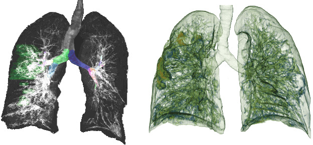
Image Classification CT ScanThis retrospective NIfTI image dataset consists of unenhanced chest CTs from 632 patients with COVID-19 infections. The images were acquired at the point of care in an outbreak setting from patients with Reverse Transcription Polymerase Chain Reaction (RT-PCR) confirmation for the presence of SARS-CoV-2.
Presented by The Cancer Imaging Archive (TCIA) Public Access,
-
COVID-Net Open Source Initiative
Mar 22, 2020 @ 11:30 PM
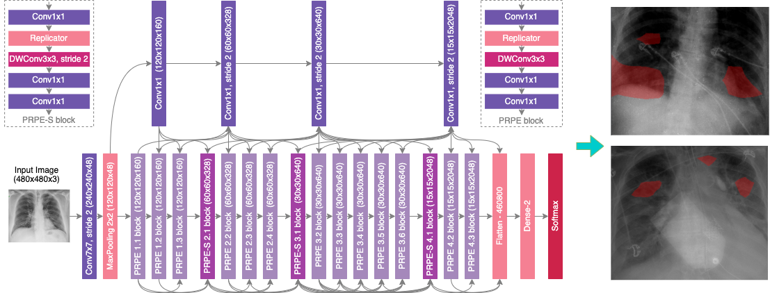
Image Classification Chest X-rayThe COVID-19 pandemic continues to have a devastating effect on the health and well-being of the global population. A critical step in the fight against COVID-19 is effective screening of infected patients, with one of the key screening approaches being radiology examination using chest radiography. It was found in early studies that patients present abnormalities in chest radiography images that are characteristic of those infected with COVID-19. Motivated by this and inspired by the open source efforts of the research community, in this study we introduce COVID-Net, a deep convolutional neural network design tailored for the detection of COVID-19 cases from chest X-ray (CXR) images that is open source and available to the general public.
Presented by DarwinAI Corp., Canada and Vision and Image Processing Research Group, University of Waterloo, Canada
-
CheXpert: A Large Chest X-Ray Dataset And Competition
Mar 22, 2020 @ 11:30 PM
Image Classification Chest X-rayCheXpert is a large dataset of chest X-rays and competition for automated chest x-ray interpretation, which features uncertainty labels and radiologist-labeled reference standard evaluation sets.
Presented by Stanford ML Group
-
ChestX-ray8: Hospital-scale Chest X-ray Database and Benchmarks on Weakly-Supervised Classification and Localization of Common Thorax Diseases
Mar 22, 2020 @ 11:30 PM
Image Classification Chest X-rayIn this paper, we present a new chest X-ray database,namely “ChestX-ray8”, which comprises 108,948 frontal-view X-ray images of 32,717 unique patients with the text-mined eight disease image labels (where each image canhave multi-labels), from the associated radiological reportsusing natural language processing. Importantly, we demonstrate that these commonly occurring thoracic diseases canbe detected and even spatially-located via a unified weakly-supervised multi-label image classification and disease localization framework, which is validated using our propose dataset.
Presented by Clinical Center. America's Research Center
-
PadChest: A large chest x-ray image dataset with multi-label annotated reports
Mar 22, 2020 @ 11:30 PM
Image Classification Chest X-rayWe present a labeled large-scale, high resolution chest x-ray dataset for automated exploration of medical images along with their associated reports. This dataset includes more than 160,000 images from 67,000 patients that were interpreted and reported by radiologists at Hospital San Juan (Spain) from 2009 to 2017, covering six different position views and additional information on image acquisition and patient demography. The reports were labeled with 174 different radiographic findings, 19 differential diagnoses and 104 anatomic locations organized as a hierarchical taxonomy mapped to standard Unified Medical Language System (UMLS) terminology
Presented by Hospital San Juan de Alicante – University of Alicante
-
VinBigData Chest X-ray Abnormalities Detection Automatically localize and classify thoracic abnormalities from chest radiographs
Mar 22, 2020 @ 11:30 PM
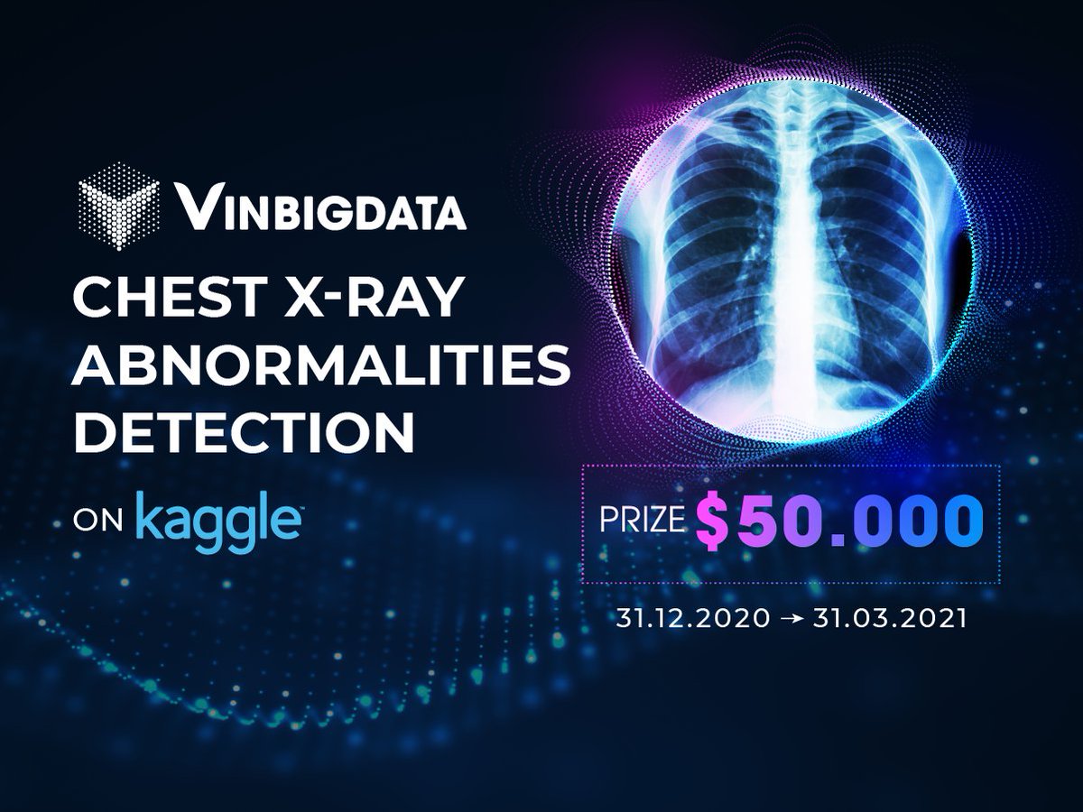
Image Classification Object Detection Chest X-rayIn this competition, we are classifying common thoracic lung diseases and localizing critical findings. This is an object detection and classification problem. For each test image, you will be predicting a bounding box and class for all findings. If you predict that there are no findings, you should create a prediction of "14 1 0 0 1 1" (14 is the class ID for no finding, and this provides a one-pixel bounding box with a confidence of 1.0). The images are in DICOM format, which means they contain additional data that might be useful for visualizing and classifying. The dataset comprises 18,000 postero-anterior (PA) CXR scans in DICOM format, which were de-identified to protect patient privacy. All images were labeled by a panel of experienced radiologists for the presence of 14 critical radiographic findings
Presented by Vingroup Big Data Institute
-
Medical Imaging Data Resource Center (MIDRC) - RSNA International COVID-19 Open Radiology Database (RICORD) Release 1b - Chest CT Covid- (MIDRC-RICORD-1b)
Jun 30, 2021 @ 11:00 PM
Image Classification CT ScanMulti-institutional, multi-national expert annotated COVID-19 imaging dataset made freely available to the machine learning community as a research and educational resource for COVID-19 chest imaging. The Radiological Society of North America (RSNA) assembled the RSNA International COVID-19 Open Radiology Database (RICORD) collection of COVID-related imaging datasets and expert annotations to support research and education. RICORD data will be incorporated in the Medical Imaging and Data Resource Center (MIDRC), a multi-institutional research data repository funded by the National Institute of Biomedical Imaging and Bioengineering of the National Institutes of Health. The RSNA International COVID-19 Open Annotated Radiology Database (RICORD) release 1b consists of 120 thoracic computed tomography (CT) scans of COVID negative patients from four international sites.
Presented by The Cancer Imaging Archive (TCIA) Public Access
-
Medical Imaging Data Resource Center (MIDRC) - RSNA International COVID-19 Open Radiology Database (RICORD) Release 1a - Chest CT Covid+ (MIDRC-RICORD-1a)
Jun 30, 2021 @ 11:00 PM
Image Classification CT ScanThe Radiological Society of North America (RSNA) assembled the RSNA International COVID-19 Open Radiology Database (RICORD), a collection of COVID-related imaging datasets and expert annotations to support research and education. The RICORD datasets are made freely available to the research community and will be incorporated in the Medical Imaging and Data Resource Center (MIDRC), a multi-institutional research data repository funded by the National Institute of Biomedical Imaging and Bioengineering of the National Institutes of Health. MIDRC-RICORD dataset 1a consists of 120 thoracic computed tomography (CT) scans from four international sites annotated with detailed segmentation and diagnostic labels.
Presented by The Cancer Imaging Archive (TCIA) Public Access
-
MosMedData: Chest CT Scans with COVID-19 Related Findings
jun 30, 2021 @ 11:00 PM
Image Classification CT ScanThis dataset contains anonymised human lung computed tomography (CT) scans with COVID-19 related findings, as well as without such findings. A small subset of studies has been annotated with binary pixel masks depicting regions of interests (ground-glass opacifications and consolidations). CT scans were obtained between 1st of March, 2020 and 25th of April, 2020, and provided by medical hospitals in Moscow, Russia..
-
COVID-19 Lung CT Lesion Segmentation Challenge - 2020
jun 30, 2021 @ 11:00 PM
Image Classification CT ScanTraining and Validation: Unenhanced chest CTs from 199 and 50 patients, respectively, with positive RT-PCR for SARS-CoV-2 and ground truth annotations of COVID-19 lesions in the lung. Testing: Additional, unseen 46 patients with positive RT-PCR for SARS-CoV-2 and ground truth annotations of COVID-19 lesions in the lung CT. The test cases are from a variety of sources, included sources not used for training and validation. CT data provided by The Multi-national NIH Consortium for CT AI in COVID-19 via the NCI TCIA public website.
Presented by Grand Challenge
